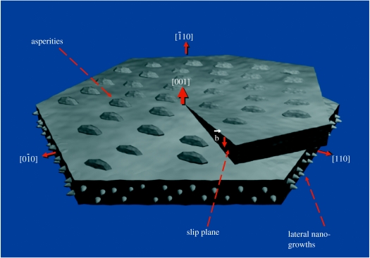
Figure 3.
A schematic of the crystallographic directions of an aragonite platelet without surrounding organic layers at a screw dislocation core and the corresponding nano-structures. Asperities, previously interpreted as mineral bridges in lower resolution images, are squat structures found on the [001] faces, while the newly observed [010] or [110] nano-growths are in the vertical plane and have a larger aspect ratio. Note the crystallographic alignment of the asperities, which indicates the platelet crystal orientation. The dislocation slip plane and Burgers vector are labelled.

