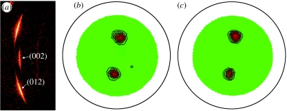
Figure 5.
(a) X-ray diffraction pattern and (b,c) pole figure texture measurements of aragonite platelets. The (002) reflection shows that the platelet surface is normal to the vertical growth direction, while the (012) reflection is used for pole figure measurement to probe the lateral orientation of aragonite platelets. The similar features presented in pole figures with both (b) 5° and (c) 10° incidence angles suggest that the preferred lateral orientation does not change with the X-ray penetration depth.

