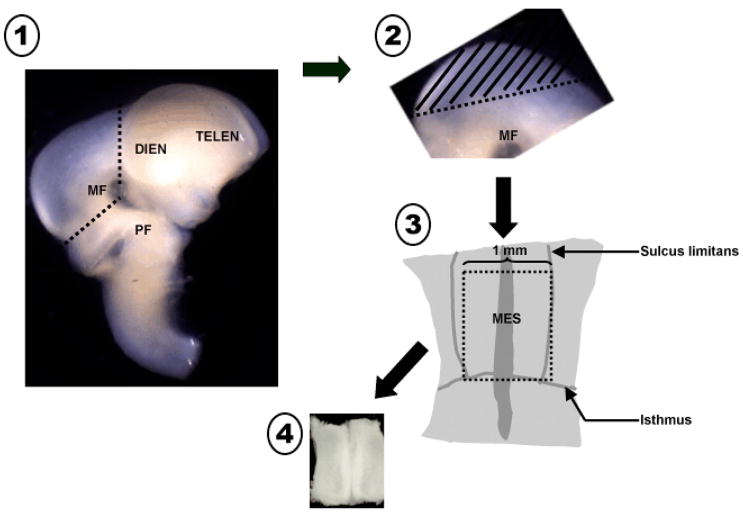
Figure 1.
Schematic diagram illustrating the microdissection technique to harvest ventral mesencephalic tissue. 1. Incisions are made at the rostral and caudal borders of the mesencephalic flexure to excise the mesencephalon. 2. The dorsal segment of the mesencephalon (tectum, //////) is removed and discarded. 3. The remaining ventral tegmental tissue is laid flat exposing the floor of the 4th ventricle. Incisions of appropriate length are made utilizing the isthmus and sulcus limitans as landmarks. 4. The isolated ventral mesencephalic tissue piece is collected and processed to single cell suspension. MF = mesencephalic flexure, PF = pontine flexure, DIEN = diencephalon, TELEN = telencephalon, MES = mesencephalon,----------- = incision line.

