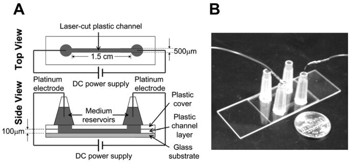
Figure 3. Microfluidic chamber for visualization of cell migration in an applied electric field.
(A) Illustration of microfluidic system for studying cell migration in an applied electric field. (B) A picture of the microfluidic system. Two identical channels were configured side-by-side in a single device and can be used for migration studies separately. A nickle was placed next to the device as a scale reference.

