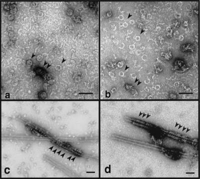
Figure 3.
DynI and dynII self-assemble into similar oligomeric structures. DynI (a) and dynII (b) were self-assembled by dialysis into HCB30 containing 1% ethylene glycol and examined by electron microscopy in negative stain. Arrowheads indicate rings and small stacks of rings. In c and d, dynI and dynII, respectively, were incubated with taxol-stabilized microtubules at 0.1 mg/ml in PH buffer. Final dynamin concentration in each condition was 0.1 mg/ml. Arrowheads show dynamin rings encircling the microtubule template. Bars, 100 nm.

