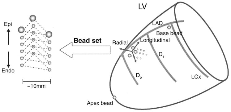
Figure 1. Schematic Representation of the Heart.
The transmural bead set was implanted between the first (D1) and the second (D2) diagonal branch of the left anterior descending coronary artery (LAD) to measure finite deformation of the myocardial tissue across the wall. Endo = endocardium; Epi = epicardium; LCx = left circumflex coronary artery; LV = left ventricle.

