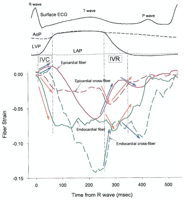
Figure 8. Transmural Tissue Coupling.
Red and green lines = strains in epicardial (0% wall depth) and endocardial (90% wall depth) layers, respectively. Solid and broken lines = fiber and cross-fiber strains, respectively. AoP = central aortic pressure; ECG = electrocardiogram; IVC = isovolumic contraction; IVR = isovolumic relaxation; LAP = left atrial pressure; LVP = left ventricular pressure.

