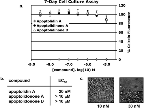
Figure 3.
Toxicity of apoptolidin A and aglycones in H292 human lung carcinoma cells. (a) Cells were plated at a density of 500/well in 96-well plates and treated for 7 days with apoptolidin A, apoptolidinone A, and apoptolidinone D at concentrations from 3 nM to 10 μM. Viability was measured by loading cells with 2 μM Calcein-AM and reading plates using a Spectramax (Molecular Dynamics) plate reader; λabs = 494, λem = 517. (b) Effective concentration 50 (EC50) values for apoptolidin A, apoptolidinone A, and apoptolidinone D. (c) Bright field photomicrographs of H292 cells cultured for 7 days in the presence of either 10 or 30 nM apoptolidin A.

