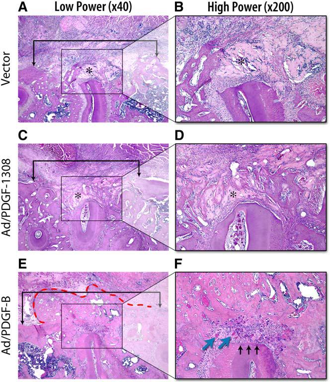
Fig. 4.

In vivo gene transfer of PDGF-B stimulates periodontal tissue regeneration. Histologic microphotographs of periodontal alveolar bone defects treated for 14 days after gene delivery of Ad/PDGF-B, Ad/PDGF-1308, or vector alone. (A, C, E, original magnification ×40; B, D, F, original magnification ×200.) Brackets in the low-power (×40) slides indicate alveolar bone wound edges, with no significant differences between the sizes of the defects based on histomorphometric analyses. Limited alveolar bone formation occurred in the Ad/PDGF-1308 and vector alone defects, whereas significant bone bridging was noted most extensively in sites treated with Ad/PDGF-B (red dashed line). (F) A thin layer of newly formed cementum (black arrows) was observed only in the Ad/PDGF-B–treated defects. More vascularization (blue arrows) was seen in the periodontal ligament region of the Ad/PDGF-B–treated lesions. Asterisks indicate the collagen carrier. (Adapted from Jin Q, Anusaksathien O, Webb SA, et al. Engineering of tooth-supporting structures by delivery of PDGF gene therapy vectors. Mol Ther 2004;9(4):522; with permission.)
