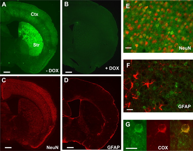
Figure 2.
Neuron-specific mitochondrial expression of eYFP and its regulation by doxycycline. A, B, Mito/eYFP expression was induced by feeding the bigenic mice with normal diet (A) or suppressed by feeding with doxycycline-containing diet (B). Coronal sections were visualized under fluorescence microscopy. C, D, Adjacent sections were counterstained with monoclonal anti-NeuN antibody (C) followed by secondary antibody (anti-mouse conjugated to Alexa Fluor 594) or with polyclonal anti-GFAP antibody (D) followed by secondary antibody (anti-rabbit conjugated to Alexa Fluor 555). Note colocalization of eYFP with NeuN staining. E, F, A higher magnification of eYFP fluorescence in cortical neurons shows colocalization with NeuN (E) but not with GFAP (F). G, Immunostaining of neurons from dentate gyrus with anti-cytochrome oxidase subunit I antibody (middle) showed eYFP expression (left) colocalized with the mitochondrial marker protein (right). Scale bars: A–D, 250 μm; E–G, 25 μm. DOX, doxycycline; Str, striatum; Ctx, cortex.

