
Figure 1.
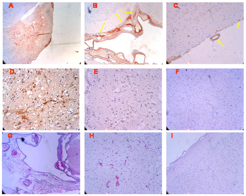
Immunohistochemistry and histopathology four hours after cerebral hypoxia/ischemia. Sections of the parietal cortex from piglet brains after ischemic injury (A-E) and from uninjured control animal (I and F), were subjected to antigen retrieval in citrate buffer and stained with anti-uPA monoclonal antibody (5 μg/ml) (Panels, A-D and F) or with non-immune mouse IgG1 as a negative control (Panel E), secondary biotinylated anti-mouse IgG (1:200), followed by incubation with HRP-conjugated streptavidin. Magnification shown is 100x for Panels A, B, G, and I, 200X for panels, C, E, F, and H, 400x for Panel D. Adjacent sections from the same brains exposed to ischemic injury (Panels G-H) and from uninjured controls (Panel I), were stained by H&E for histological inspection. These data reflect an n of 2 per experimental group.