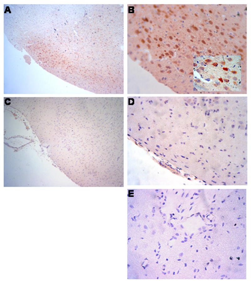
Figure 6.

Immunhistochemistry for phosphorylation of ERK MAPK four hours after cerebral hypoxia/ischemia. Sections of the parietal cortex from piglet brains after ischemic injury (A,B, and E) and from uninjured control animal (C and D) were stained with phospho-p44/42 MAPK rabbit monoclonal antibody (Panels A-D) or with rabbit IgG as a negative control (Panel E). Magnification shown is 100X for Panels A and C, 400X for Panels D,B, and E, X1000 for insert in Panel B. These data reflect an n of 2 per experimental group.
