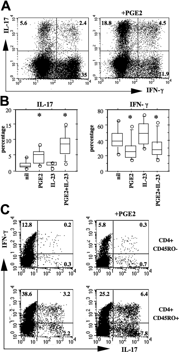
Figure 3.

PGE2 and IL-23 favor Th17 expansion. (A,B) Culture conditions as in the legend of Figure 1. Cells were harvested after 7 days of culture and processed for intracellular staining on PMA and ionomycin activation as described in “Flow cytometry.” (A) Representative example of a culture in the presence of IL-23 (10 ng/mL) with or without PGE2 (50 ng/mL). (B) Box plots represent the 10th, 25th, 50th, 75th, and 90th percentiles of 7 distinct individual donors. Circles represent outliers. *Significant differences (P < .01) compared with the nil culture condition by paired Student's t test. Note that IL-17 positivity was restricted to CD4+ T cells. (C) Peripheral blood CD4+ T cells were sorted into CD45RO− (naive) and CD45RO+ (memory) subsets, activated by CD3/CD28-coated beads, and cultured in the presence of IL-2 (20 U/mL), IL-23 (10 ng/mL), with or without PGE2 (50 ng/mL). Experiment representative of similar results with cells from three distinct donors.
