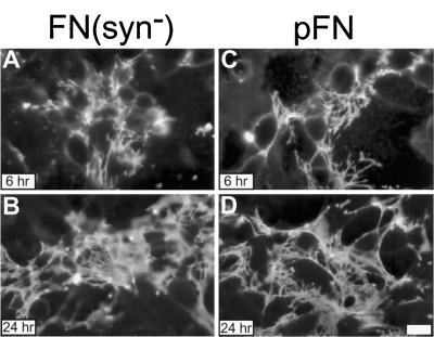
Figure 3.
FN(syn−) and pFN matrix assembly supported by αvβ3. αvβ3-transfected CHO K1 cells were incubated with 20 μg/ml cycloheximide to inhibit endogenous FN synthesis, monoclonal antihamster α5 integrin function blocking antibody, activating LIBS6 anti-β3 antibody, and 25 μg/ml of either FN(syn−) (A and B) or pFN (C and D) for 6 and 24 h. FN fibrils were visualized by indirect immunofluorescence using monoclonal antibody IC3. Bar, 10 μm.

