Fig. 1.
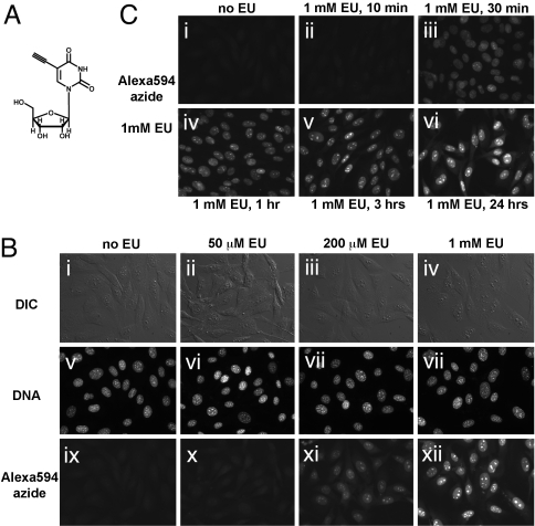
Imaging cellular transcription by using EU. (A) Structure of the uridine analog EU, a biosynthetic RNA label. (B) EU incorporation into RNA in NIH 3T3 cells. Cells were grown without EU (i, v, and ix), with 50 μM EU (ii, vi, and x), 200 μM EU (iii, vii, and xi), or 1 mM EU (iv, viii, and xii) for 20 h. The cells were fixed and reacted with 10 μM Alexa594-azide. EU-labeled cells show strong nuclear and weaker cytoplasmic staining, proportional to the added EU concentration. Note the intense staining of nucleoli. All cells incorporate EU, although some cell-to-cell variability is observed. (C) Rapid uptake and incorporation of EU by cells. NIH 3T3 cells were incubated with 1 mM EU for varying amounts of time, followed by fixation and EU detection. Strong nuclear staining is visible after 30 min (iii), although even after 10 min (ii), a signal above background is observed at longer exposure times (data not shown). EU staining intensity increases quickly in the first 3 h (iv and v) and then more slowly up to 24 h (vi).

