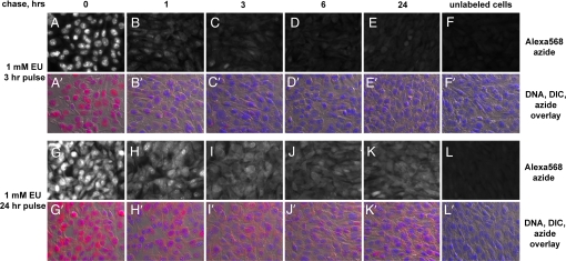
Fig. 4.
Imaging cellular RNA turnover with EU. NIH 3T3 cells were labeled for 3 h (A–F and A′–F′) or 24 h (G–L and G′–L′) with 1 mM EU. The label was chased with complete media for different periods of time. The cells were fixed and stained with Alexa568-azide and Hoechst. After a 3-h pulse the nuclear EU staining drops quickly in the first hour of the chase (A and B; A′ and B′) and becomes very low after 6 h (D and D′). EU staining of the cytoplasm shows a delayed decline compared with the nuclear signal. Cytoplasmic staining is still visible after 6 h (D and D′), whereas after 24 h (E and E′) it drops very close to background levels (F and F′). After labeling with EU for 24 h, both the nucleus and the cytoplasm are strongly labeled (G and G′). The nuclear signal drops significantly during the chase but does not disappear. Strong cytoplasmic staining persists after a 24-hour chase, indicating the labeling of stable RNA species.

