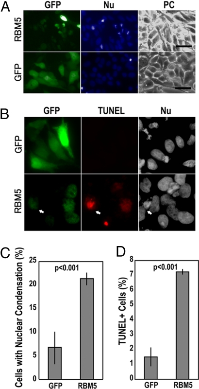
Fig. 3.
Induction of cell death by RBM5 overexpression. (A and B) Micrographs of GFP, nuclear morphology (Nu), or phase-contrast image (PC), or TUNEL staining of HeLa cells transfected with GFP-tagged RBM5 or GFP-vector control. Cell death was examined by nuclear staining with Hoechst dye (A) and TUNEL staining (B). (Scale bar: 50 μm.) (C and D) Quantification of cell death in transfected cells as determined by the percentages of cells showing condensed or fragmented nuclei (C) or cells showing positive TUNEL staining (D; see Materials and Methods). The arrow in B marks a typical GFP-RBM5-expressing cell that shows positive TUNEL staining. We noticed that many GFP-RBM5-positive cells undergoing apoptosis became detached during washing procedures in the TUNEL staining, providing an explanation of the overall low level of TUNEL-positive cells photographed and scored. Approximately 1,000 cells were examined in each group, and the data represent three independent experiments.

