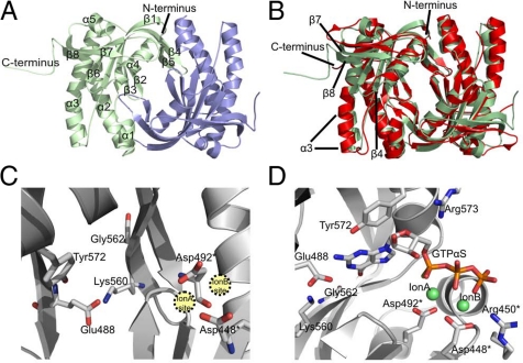
Fig. 3.
Crystal structure of Cya2 catalytic domain. (A) Overall structure of the Cya2 homodimer. The two monomers are colored green and blue, and the secondary structure elements are labeled in one monomer. (B) Overlay of the Cya2 catalytic domain (green) with the AC enzyme CyaC (red). Secondary structure elements displaying differences between the two cyclases are labeled. (C) Active site of the Cya2 dimer. Residues mentioned in the text are labeled; residues supplied by the second monomer of the dimer are marked with an asterisk. (D) Cya2 active site with a modeled GTPαS ligand. The nucleotide was positioned in Cya2 after superposition with the structure of a CyaC/ATPαS complex; the nucleotide base was then modified manually to guanosine.

