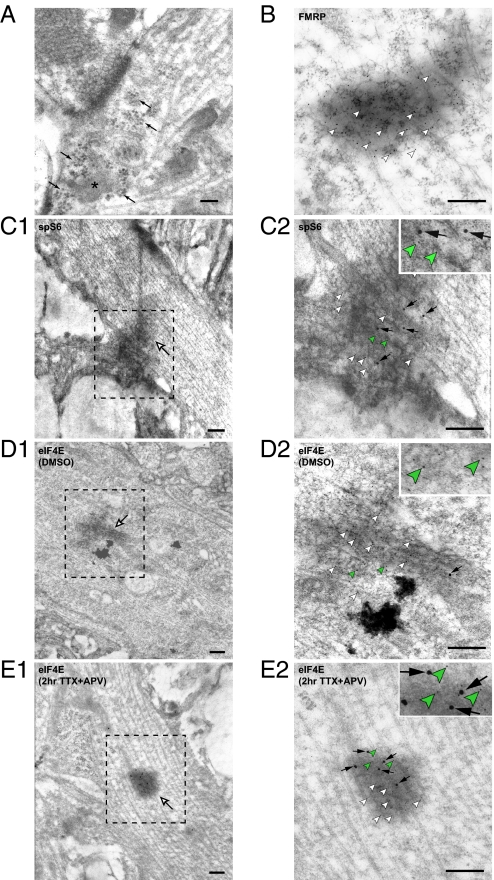
Fig. 3.
Characterization of neuronal RNA granules. (A) Polyribosomes (black arrows) are present in neuronal dendrites. RNA granule-like electron-dense dark complexes lacking a clear membranous boundary (asterisk) are sometimes associated with polyribosomes. (Scale bar: 100 nm.) (B) FMRP protein (5-nm gold, white arrowheads) is enriched in these dendritic dark complexes. (Scale bar: 200 nm.) (C1) Ribosomal protein S6 (15-nm gold) is present in dendritic RNA granules (white arrow). (Scale bar: 200 nm.) (C2) The image of the RNA granule in C1 under higher magnification. The 5-nm (white and green arrowheads) and 15-nm gold particles (black arrows) label FMRP and S6 respectively. (Scale bar: 200 nm.) (D–E) Double immunolabeling of FMRP (5-nm gold, white and green arrowheads) and eIF4E (15-nm gold, black arrows in D2 and E2) in dendritic RNA granules (white arrows in D1 and E1). (D1) The eIF4E protein level in RNA granules is undetectable under basal conditions. (D2) The image of the RNA granule in D1 at higher magnification. (E1) eIF4E is recruited into RNA granules (black arrow) upon translation activation by treatment with TTX and APV. (E2) The image of the RNA granule in E1 at higher magnification. (Scale bars: D–E, 200 nm.)

