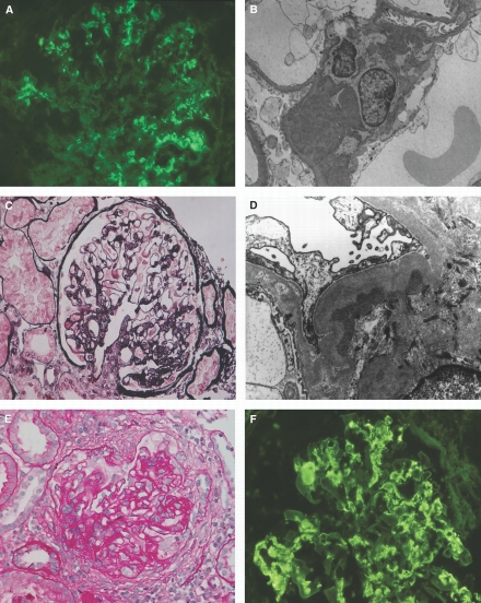
Figure 1.
Pathologic findings in C1q nephropathy. (A and B) A 14-yr-old male patient presenting with asymptomatic hematuria and proteinuria. (A) Immunofluorescence shows global granular mesangial staining for C1q. (B) Electron micrograph displays dense deposits in the mesangium without cell proliferation. (C and D) A 27-yr-old female patient presenting with nephrotic syndrome. (C) A glomerulus displays segmental perihilar glomerular sclerosis, whereas most of the glomeruli (data not shown) were unremarkable (periodic acid silver methenamine/Azan). (D) On electron microscopy, dense deposits are seen in the widened mesangial matrix beneath the glomerular basement membrane. Note also extensive foot process effacement and segmental podocyte-free surface microvillous transformation. (E and F) A 29-yr-old male patient presenting with chronic kidney disease. (E) A glomerulus shows mesangial and endocapillary proliferation accompanied by extensive fibrocellular crescent (periodic acid Schiff). (F) Immunofluorescence shows conspicuous mesangial and segmental capillary wall C1q staining in “full house” pattern glomerular immune deposits. Magnifications: ×400 in A, C, E, and F; ×4400 in B; 10,000 in D.

