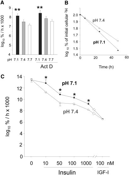
Figure 3.
(A) Effect of 1 μM Actinomycin D (Act D) on proteolysis rate in L6-G8C5 myotubes during 7-h incubations at the specified pH in MEM + 2 mM L-Phe + 2% dialyzed FBS. Pooled data from four independent experiments are shown (with three replicate culture wells for each treatment). Proteolysis was assessed from the rate of release of radioactivity into the medium from cultures prelabeled with 3H-L-Phe and is expressed as the logarithm of the percentage of the total initial cellular radioactivity per hour (log10 %/h × 1000; see the Concise Methods section). (B) Typical delabeling time course of 3H-L-Phe–prelabeled cells incubated in serum-free MEM + 2 mM L-Phe at the specified pH with 100 nM insulin. Linear regression slopes of plots of this type are presented in A and C and Figure 4. (C) Effect of pH, insulin, and high-dosage IGF-1 on the rate of proteolysis (delabeling) expressed as log10 %/h × 1000 in L6-G8C5 myotubes. Pooled data from three independent experiments are shown (with three replicate culture wells for each treatment). *P < 0.05 versus the corresponding pH 7.4 control value; **P < 0.05 versus the corresponding pH 7.7 value.

