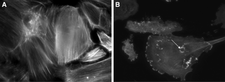
Figure 1.
(A) WT podocytes exhibited actin in stress fibers in normal culture conditions and in SFM. (B) By contrast WT podocytes treated with Hx (0.05 mg/ml for 30 min) showed dramatic reorganization of actin with loss of stress fibers and peripheral ruffles and the development of cytoplasmic aggregates. In some cells, these aggregates resembled podosomes (arrow). Images are representative of n = 6 independent experiments.

