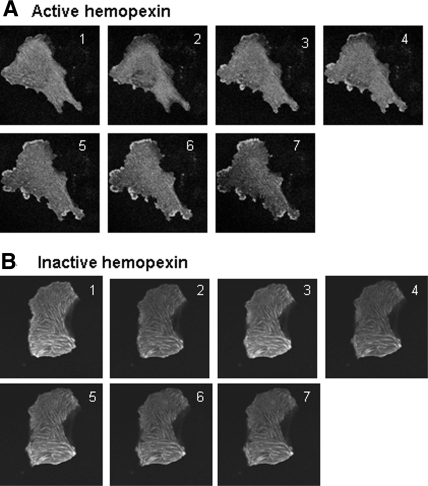
Figure 3.
(A and B) Real-time, laser scanning, confocal microscopy images of WT podocytes microinjected with GFP actin and then treated with active Hx (A) and heat-inactivated Hx (B) (0.02 to 0.05 mg/ml for 30 min). With active Hx, there was a dramatic reorganization of the actin cytoskeleton, with the development of membrane ruffles and cytoplasmic actin aggregates and the loss of actin stress fibers. Representative of n = 3 independent experiments performed in duplicate. Selected stills from a typical experiment are shown (complete animation of this experiment can be seen as supplemental information online).

