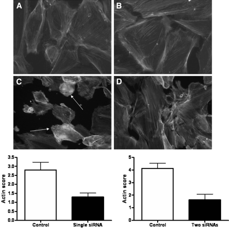
Figure 4.
(A) ND podocytes exhibited actin in stress fibers in normal culture conditions and in SFM. (B) Unlike WT podocytes, ND podocytes did not show actin reorganization after treatment with Hx (0.05 to 0.10 mg/ml for 30 min). Images are representative of n = 4 independent experiments. (C and D) Furthermore, siRNA knockdown of nephrin in WT podocytes followed by treatment with Hx (0.05 to 0.10 mg/ml for 30 min) was associated with a reduction in actin reorganization (D) compared with control siRNA and Hx treatment (C). Arrows indicate actin reorganization. The changes were assessed with an actin scoring system by two independent observers, and there were significant differences (P < 0.05) using both single and two siRNA sequences as shown in the graphs.

