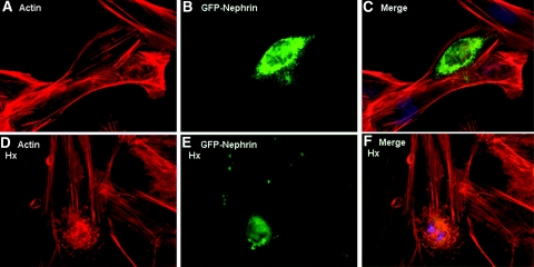
Figure 5.
ND podocytes were reconstituted with GFP-tagged nephrin by transient transfection. (A through C) Cells expressing GFP-nephrin (B) demonstrated actin stress fibers in SFM (A and merged in C). (D through E) When these cells were treated with Hx, (0.05 to 1.00 mg/ml for 30 min), only the GFP-nephrin–expressing cells (E) demonstrated actin reorganization (D and merged in F). Images are illustrative of n = 3 experiments each performed in triplicate.

