Figure 3.
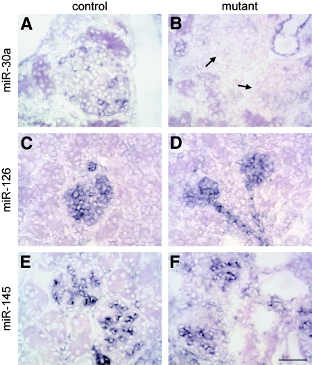
miRNA processing is abolished in mutant podocytes. miRNA expression was evaluated in the kidneys of 3-wk-old by in situ hybridization. (A) miR-30a was expressed by collecting duct epithelium and podocytes in controls. (B) Podocytes in mutant glomeruli (arrows) were negative, a finding consistent with loss of Dicer activity, whereas tubular labeling was comparable to controls. This involved all podocytes, including those populating superficial glomeruli that show no pathology. (C and D) Signals for miR-126 were detected in glomerular and peritubular capillary endothelial cells in controls (C), and labeling was comparable in mutants (D). (E and F) miR-145 was expressed by mesangial and vascular smooth muscle cells in normal kidney (E), and labeling was indistinguishable in mutants (F). Bar = 50 μm; n ≥ 4 mice for all panels.

