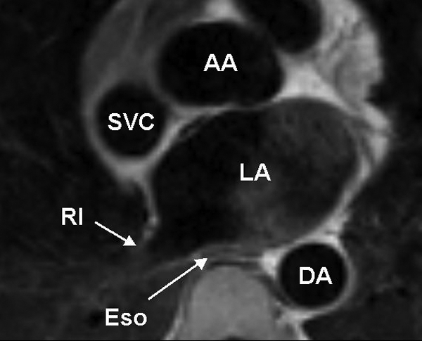
Figure 1.

Axial spin-echo T1 weighted cardiovascular magnetic resonance at the level of the left atrium (LA) and right inferior pulmonary vein (RI). The oesophagus (Eso) is immediately posterior to the left atrium and adjacent to the right inferior pulmonary vein. The oesophagus is compressed between the enlarged left atrium and the spinal column. The ascending aorta (AA), descending aorta (DA), and superior vena cava (SVC) are also shown.
