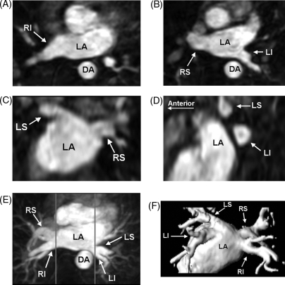
Figure 2.
Pulmonary vein three-dimensional contrast-enhanced magnetic resonance angiography showing the normal anatomy and quantification of pulmonary vein size. These images show the normal complement of four pulmonary veins, along with left atrium (LA) and descending aorta (DA). The right inferior (RI), right superior (RS), and left inferior (LI) pulmonary veins are shown in the axial plane (A and B). The left superior (LS) and right superior pulmonary veins are shown in the coronal plane from the posterior–anterior orientation (C). The left superior (LS) and left inferior (LI) pulmonary veins are shown in the sagittal plane (D, anterior to the left). All of the pulmonary veins are shown in the axial maximal intensity projection (E) and posterior–anterior volume rendered (F) image. The aorta has been removed from the volume rendered image to show all of the pulmonary veins. All of these images were derived from the same three-dimensional magnetic resonance angiography dataset. The gray lines in the maximal intensity projection image (E) correspond to the location in the sagittal plane in which the pulmonary veins separate from the left atrium and from each other. The maximal diameter, perimeter and cross-sectional area can be easily measured in the sagittal images.

