Figure 4.

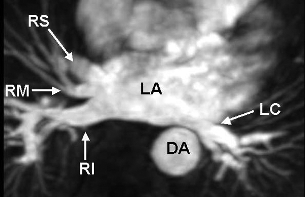
Three-dimensional contrast-enhanced magnetic resonance angiography of variant pulmonary venous anatomy. This image was obtained from a patient with right middle (RM) and left common (LC) pulmonary veins.

Three-dimensional contrast-enhanced magnetic resonance angiography of variant pulmonary venous anatomy. This image was obtained from a patient with right middle (RM) and left common (LC) pulmonary veins.