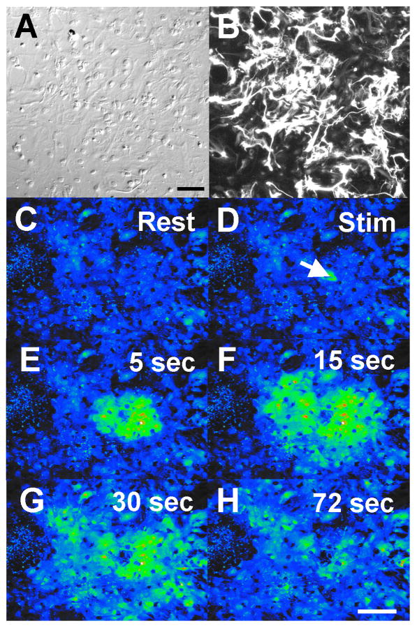
Figure 1.
Ca+2 wave propagation through a confluent mammalian astrocyte culture. A-B. DIC image of a region of interest of telencephalic mammalian astrocytes (A). The same field of view as in A is illustrated for anti-GFAP fluorescence (B). Astrocyte cultures were 85% glial as determined by GFAP immunocytochemistry. Bar equals 100 μm (for panels A-B). C-H. Pseudo-colored image of an intercellular calcium wave. A region of telencephalic mouse astrocytes, loaded with Fluo-4 AM, shows selected frames from a calcium wave sequence. After resting levels of calcium were obtained, a single cell was activated by mechanical stimulation (arrow in D; Stim) and this touch stimulation initiated a Ca+2 wave that spread to surrounding glial cells. Bar equals 200 μm (for panels C-H).

