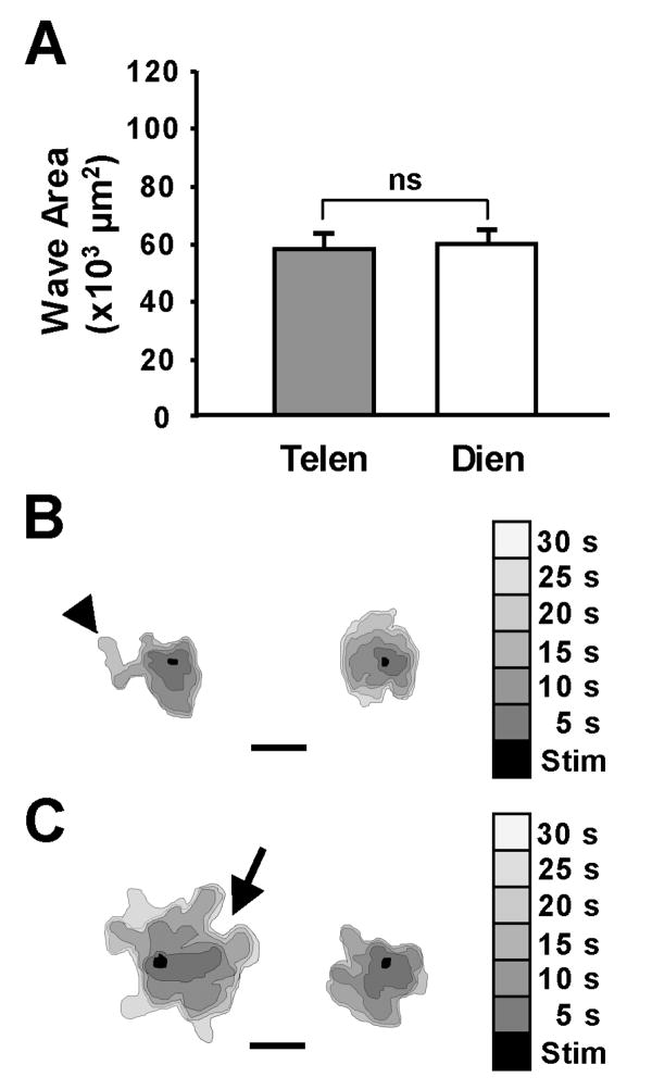
Figure 3.
The spread of calcium waves among avian astrocytes was homogeneous. A. In chick astrocyte cultures, the spread of the Ca+2 waves after 90 seconds was the same in telencephalic (Telen; n=12) and diencephalic (Dien; n=27) cultures. However the spread of the waves was dramatically reduced from those seen in the mammalian astrocytes. Note the scale of the y-axis. B-C. The nature of the spread of the wave was similar in both the chick telencephalic (B) and diencephalic (C) cultures. Two representative waves from each group indicate the area of wave spread in 5-second intervals (shades of gray) from the touch stimulation site (black area). Calcium waves among the astrocyte cultures made hairpin turns (arrow) and had unequal advances of the wave front (arrowhead). Bar equals 200 μm.

