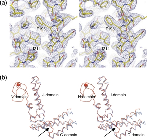
FIGURE 1.
Three-dimensional structure of hHscB. a, stereodiagram of a representative portion of the 3.0 Å 2mFo-DFc electron density map (blue mesh) of hHscB and final refined model (sticks). b, stereodiagram of Cα traces of structurally superposed hHscB (red) and E. coli HscB (cyan). The structure of hHscB consists of three domains: the N-domain, which is not found in E. coli HscB, and the J- and C-domains connected by a short linker region (black arrow). The red sphere represents a metal ion coordinated by the N-domain.

