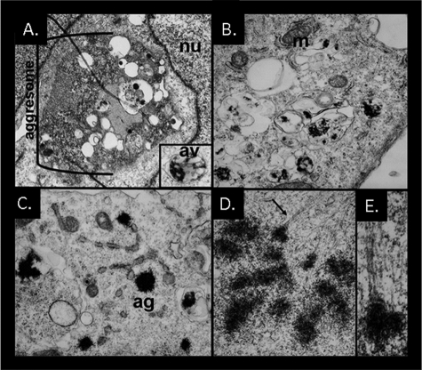
FIGURE 3.
Aggresome formation is impaired in IBMPFD mutant-expressing cells and tissue. Electron micrographs of U20S cells co-expressing p97/VCP-WT (A) or p97/VCP-R155H (B-E) along with polyQ80-CFP for 48 h. A, p97/VCP-WT-expressing cells have large perinuclear inclusions with active autophagic degradation consistent with an aggresome. The inset shows a double membrane containing autophagic vacuole with electron dense debris. B and C, IBMPFD mutant-expressing cells have multiple small electron dense inclusions that are within membranous structures or non-membrane-associated within the cytoplasm. D and E, an IBMPFD mutant-expressing cell with multiple inclusions attached to microtubules. Similar results were also obtained with p97/VCP-A232E-expressing cells.

