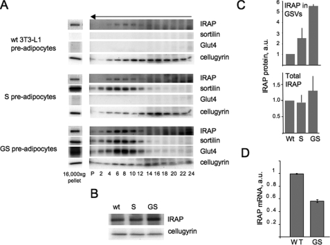
FIGURE 4.
Recruitment of IRAP into the vesicular fraction in undifferentiated 3T3-L1 cells. A, wild type (wt) 3T3-L1 cells as well as 3T3-L1 cells stably expressing sortilin (S cells) and sortilin together with Glut4 (GS cells) were homogenized and centrifuged at 16,000 × g for 20 min. Pellets of this centrifugation (30 μg) were separated by PAGE, and proteins were detected on the same membrane by sequential rounds of Western blotting. Supernatant (200-400 μg in different experiments) was centrifuged in a 10-30% linear sucrose gradient for 65 min in a Sorvall TST60.4 rotor at 48,000 rpm. The arrow indicates the direction of sedimentation. Gradient fractions, including the pellet of this centrifugation (P) were separated by PAGE. IRAP, sortilin, Glut4, and cellugyrin from each cell line were detected on the same membrane by sequential rounds of Western blotting. B, total protein lysates of wild type, G, and GS pre-adipocytes (50 μg each) were analyzed by Western blotting. C, quantification of data shown in panels A and B (mean values ± S.E. of two (S cells) or three (wild type and GS cells) independent experiments). a.u., arbitrary units. D, the levels of IRAP mRNA were determined in wild type and GS pre-adipocytes by quantitative real time PCR and normalized by glyceraldehyde-3-phosphate dehydrogenase mRNA.

