FIGURE 5.
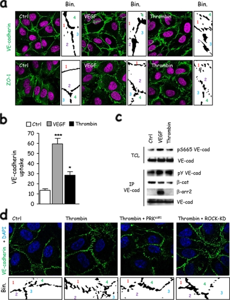
ROCK- and PRK-induced contractility is involved in cell-cell contact remodeling. a, human endothelial cells were grown on collagen-coated glass coverslips for 3 days as monolayers, starved overnight, and then stimulated with thrombin and VEGF. Cell-cell junctions (green) were revealed by either VE-cadherin or ZO-1 immunostaining. Nuclei were counterstained by 4′,6-diamidino-2-phenylindole (DAPI)(magenta). Binary calculation was applied on threshold pictures to unveil cell-cell morphology (ImageJ software). Individual cells are numerated 1–4. b, human endothelial cells were grown on collagen-coated glass coverslips for 3 days as monolayers, starved overnight, incubated with anti-VE-cadherin at 4 °C for 1 h, and then stimulated with thrombin and VEGF at 37 °C for 30 min. Cells were subjected to a mild acid-wash prior to fixation and immunofluorescence. Cells with internal acid-resistant staining for VE-cadherin were scored positive when they displayed at least five vesicle-like internal staining. Graph represents the percentage of cells with VE-cadherin uptake in the total cell population. c, human endothelial cells were grown for 3 days as monolayers, starved overnight, and then stimulated with thrombin and VEGF. Total cell lysates (TCL) were blotted against phospho-Ser-665 VE-cadherin and total VE-cadherin (VE-cad). Phosphotyrosine (pY), β-catenin (β-cat), β-arrestin2 (β-arr2), and VE-cadherin Western blots were performed in VE-cadherin immunoprecipitation (IP) fractions. d, mock-, PRK siRNA-, and ROCK-KD-transfected human endothelial cells were grown on collagen-coated glass coverslips for 3 days as monolayers, starved overnight, and then stimulated with thrombin. VE-cadherin immunostaining is shown in green. Nuclei were counterstained by 4′,6-diamidino-2-phenylindole (DAPI)(blue). Binary calculation (bin.) was applied on threshold pictures to unveil cell-cell morphology (ImageJ software). Individual cells are numerated 1–4. All panels are representative of at least three independent experiments. ANOVA test: *, p < 0.05; **, p < 0.01; ***, p < 0.001.

