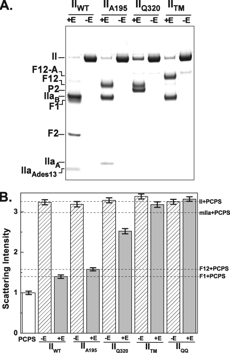
FIGURE 1.
Prolonged action of prothrombinase on prothrombin variants. The indicated prothrombin variants (3 μm) were incubated at 25 °C for 30 min with 30 nm Va, 50 μm PCPS (-E), or 3 nm Xa, 30 nm Va, 50 μm PCPS (+E). A, prothrombin cleavage was assessed by SDS-PAGE following disulfide bond reduction with bands visualized following staining with Coomassie Brilliant Blue. The various fragments are identified in the left margin. B, right angle light scattering intensities measured for the same samples diluted as described under “Experimental Procedures.” Scattering intensities were normalized by setting the signal observed with 50 μm PCPS to 1. Error bars (±2 S.D.), the variation in signal over a 3-min measurement period. Because light scattering intensity is highly dependent on experimental conditions, the error bars provide appropriate confidence limits for assessing changes within an experiment but are probably inaccurate in describing variation between experiments done on different days. Intensities measured with the indicated purified proteins (1.4 μm) plus 50 μm PCPS are indicated by the dashed lines and in the right margin.

