FIGURE 8.
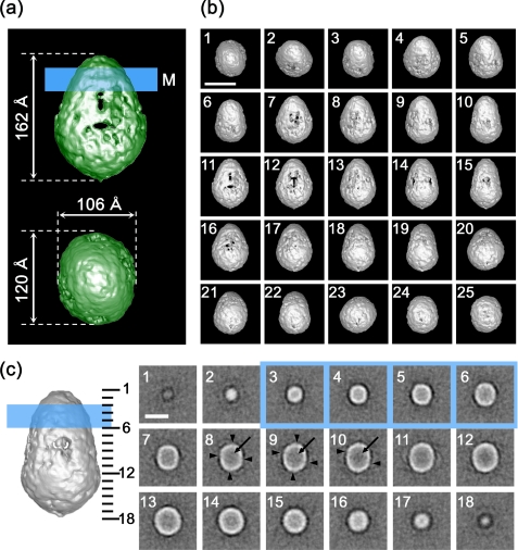
Surface representation of CFTR. a, side and top views of the CFTR protein. CFTR presumably in a closed state is shown to be an ellipsoidal particle with dimensions of 120 × 106 × 162 Å. The molecular mass enclosed by the isosurface is 276 kDa, corresponding to 81.7% of the dimeric CFTR protein. The putative position of the plasma membrane is indicated on the side view by a blue line of 30 Å in thickness (M) so that the ratio of each domain is close to the prediction from the amino acid sequence. b, surface views from 25 different angles. c, sections normal to the symmetric axis at 9.5-Å intervals through the molecule. The number in each panel corresponds to the slice position indicated on the narrower side view (left). Slices included in the transmembrane domain are marked by blue. Rifts in the cytoplasmic surface (arrowheads in panels 8-10) correspond to the orifices in the surface representation. The inside of the molecule is mostly low density (arrows), suggesting that stain permeates into the molecule. The scale bars represent 100 Å.

