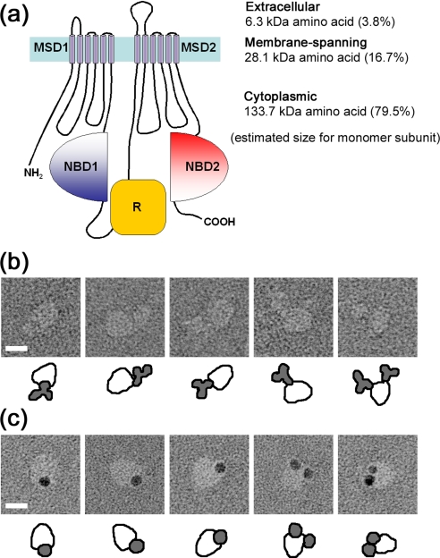
FIGURE 9.
Binding of anti-R domain antibody to CFTR. a, transmembrane topology predicted for CFTR. The subunit is composed of two repeated motifs, each of which comprises an MSD and a cytoplasmic NBD. An R domain is located between NBD1 and MSD2. The molecule is divided into 3.8% extracellular, 16.7% membrane-spanning, and 79.5% cytoplasmic domains, calculated from the amino acid sequence. b, gallery of negatively stained CFTR·anti-R domain antibody (MAB1660) complexes. MAB1660 specifically binds to the larger domain of CFTR. c, gallery of CFTR·MAB1660·protein G-gold complexes. Protein G-gold was mixed with CFTR·MAB1660 complexes on the WGA column, and then excessive gold was washed out. Similar to the observation in b, the protein G-gold again binds to the larger domain. CFTR particles bearing two gold particles were also observed. The scale bars represent 100 Å.

