Figure 1.
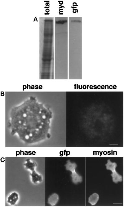
(A) Expression of GFP-myosin in myosin-null Dictyostelium cells. Whole-cell lysates were run on SDS-PAGE. Lane 1, Coomassie stained; lane 2, antimyosin antibody; lane 3, anti-GFP antibody. A single band was identified by both antibodies, suggesting that the only GFP-labeled protein in the cell was myosin. The lighter band in the myosin blot is an antigen recognized by the secondary antibody and is not a myosin. (B) Minimal background fluorescence is seen in the myosin-null parent cell line. Live myosin-null cells were imaged with the same exposures used to image the GFP-myosin-containing cells. The cell shown in phase on the left has only minimal fluorescence as shown on the right. Bar, 3 μm. (C) GFP and myosin colocalize in vivo. A dividing GFP-myosin cell was fixed and stained with an antibody against myosin. The immunofluorescence localization of myosin mirrors that of the GFP fluorescence, implying that fluorescence localization is a reasonable marker of GFP-myosin localization. Bar, 10 μm.

