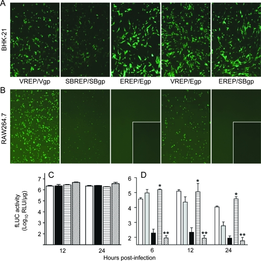
FIG. 5.
The restricted tropism of EEEV for myeloid lineage cells is not mediated by the virus-receptor interaction. (A and B) Cells of the mesenchymal (A) (represented by BHK-21 fibroblasts) or myeloid (B) (represented by RAW 264.7 monocytes/macrophages) lineage were infected at equal multiplicities (MOI = 1) with GFP-expressing parental or chimeric replicon particles and photographed using a fluorescence microscope to reveal GFP-positive cells. Replicons are as follows: VEEV replicon genome with VEEV structural proteins (VREP/Vgp) or EEEV structural proteins (VREP/Egp), SB replicon genome with SB structural proteins (SBREP/SBgp), and EEEV replicon genome with EEEV structural proteins (EREP/Egp) or SB structural proteins (EREP/SBgp). Inset panels for EREP/Egp and EREP/SBgp depict infection at 100-fold-higher MOIs than those of other infections. (C and D) Parallel experiment in which fibroblasts (C) or CD-1-derived cDCs (D) were infected with fLUC-expressing parental and chimeric replicons: VREP/Vgp (white bars), SBREP/SBgp (gray bars), EREP/Egp (black bars), VREP/Egp (horizontally hatched bars), and EREP/SBgp (diagonally hatched bars). Cells were harvested for luciferase activity assay at various times p.i. Values represent the geometric mean fLUC activities (log10 RLU/μg). Datum points are shown ± standard deviations where n = 3. For panel D, a single asterisk indicates that VREP/Egp is not significantly reduced compared with VREP/Vgp (P > 0.5) and a double asterisk indicates that EREP/SBgp is not significantly increased versus EREP/Egp (P > 0.5); EREP/Egp values are significantly reduced compared with those for SBREP/SBgp and VREP/Vgp at all time points (P < 0.01).

