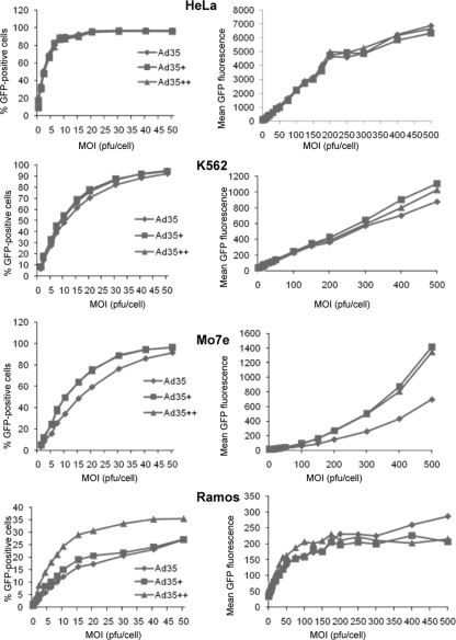
FIG. 5.
Transduction of human cell lines. Cells were infected with Ad5/35, Ad5/35+, and Ad5/35++ at the indicated MOIs, and the percentages of GFP-expressing cells (left) and GFP mean fluorescence (right) at 24 h were measured by flow cytometry. Shown are the averages of three independent experiments. The standard deviations were less then 10% from the averages for all data points.

