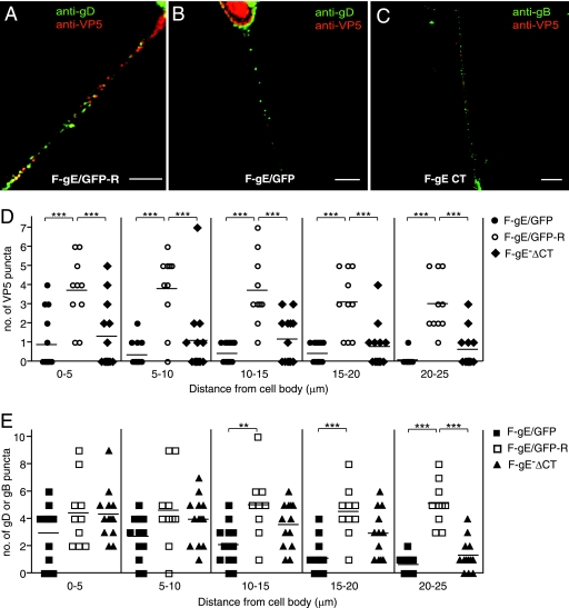
FIG. 2.
Axonal transport of capsids and glycoproteins in human neurons infected with HSV gE mutants. SK-N-SH neurons were infected with the repaired F-gE/GFP-R (A), the gE-null mutant F-gE/GFP (B), or a gE mutant lacking the gE CT domain, F-gEΔCT (C), for 18 h; fixed; permeabilized; and stained with mouse anti-VP5 MAb and rabbit anti-gD polyclonal antibodies (A and B) or mouse anti-VP5 MAb and rabbit anti-gB polyclonal antibodies (C), followed by Texas red-conjugated donkey anti-mouse and Cy-5-conjugated anti-rabbit secondary antibodies. Scale bars, 5 μm. Shown is the quantification of VP5 puncta (D) and gB or gD puncta (E) in segments of neuronal axons (0 to 5, 5 to 10, 10 to 15, 15 to 20, or 20 to 25 um) measured from the neuronal cell body. Puncta numbers were obtained from 13 F-gE/GFP-infected, 10 F-gE/GFP-R-infected, and 13 FgE ΔCT-infected neurons involving four independent experiments. Each symbol represents the number of VP5 or glycoprotein puncta observed in the indicated axon segment. Statistically significant values are shown as asterisks, with a P value of <0.05 marked as two asterisks and a P value of <0.001 marked as three asterisks.

