FIG. 7.
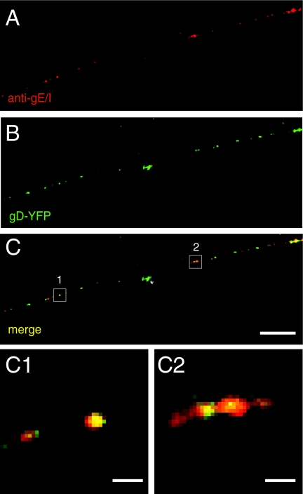
gE/gI associates with glycoprotein gD in axons of HSV-infected neurons. SK-N-SH neurons were infected with SC16 gD-YFP for 24 h, fixed, permeabilized, and stained with rat anti-gE/gI polyclonal antibodies, followed by Texas red-conjugated donkey anti-mouse secondary antibodies. Representative panels of four independent experiments are shown. (A) Rat anti-gE/gI staining. (B) SC16-gD-YFP fusion protein fluorescence. (C) Merged fluorescence. (C1 and C2) Higher magnifications of the boxed areas in panel C. Scale bars, 5 μm (A to C) and 1 μm (C1 and C2). The star indicates gD-YFP fluorescence associated with a particle that was not present within the axon shown.

