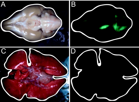
FIG. 4.
Macroscopic visualization of eGFP expression in brain and lungs of an animal that succumbed to infection with 58utrMF-NP virus. The contours of the organs are outlined by a white line. (A and B) Ventral view of the brain at the time of euthanasia (28 d.p.i.). (C and D) Ventral view of the lungs at the time of euthanasia (28 d.p.i.). Organs were photographed using phase contrast (A and C) and fluorescence excitation (B and D).

