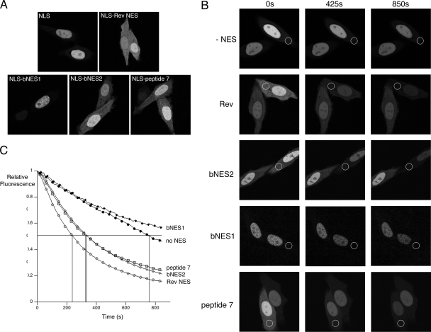
FIG. 3.
hUL47 contains a discrete NES at its extreme C terminus. (A) Hep-2 cells grown in glass-bottomed dishes were transfected with plasmids encoding GFP-NLS fused to the NES sequences from HIV-1 Rev, bUL47 (bNES1 and bNES2), and hUL47 (peptide 7). Representative images were acquired 20 h later using confocal microscopy. (B) Hep-2 cells transfected with the plasmids used for panel A were subjected to FLIP analysis as described for Fig. 1A, except that bleaching repeats were performed every 8 s. Photobleached areas are indicated by white circles. (C) The fluorescence intensity in the nuclei of bleached cells was quantified for each time point using LSM software, and relative fluorescence was calculated and plotted against time using Kaleidagraph software. The data are averages of three FLIP analyses for each fusion protein.

