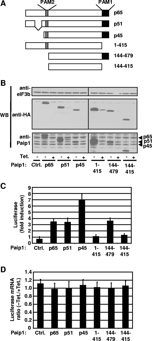
FIG. 6.
Paip1-dependent translation stimulation in vivo. (A) Schematic representation of HA-tagged Paip1 fragments expressed in HeLa cells. (B) Cells were transfected with the indicated pTet-HA-Paip1 plasmids together with constructs expressing the Renilla luciferase and the tTA. Cells were placed in medium containing 0 or 300 ng/ml of Tet. Extracts were subjected to SDS-PAGE and Western blotting (WB) with the indicated antibodies. (C) Renilla luciferase activity was quantified in HeLa cell extracts from panel B and normalized on the total protein level. Relative induction of the luciferase reporter was determined by calculating the ratio of Renilla luciferase activity between induced (without Tet) and repressed (300 ng/ml Tet) expression of the indicated HA-tagged Paip1. Error bars denote the standard error of the mean for three independent experiments. (D) Renilla luciferase mRNA was measured by quantitative RT-PCR from total RNA extracted from duplicate HeLa cells from panel B. Total RNA was DNase-treated and reverse transcribed. Using RT products, Renilla mRNA was quantified and normalized against β-actin mRNA. Luciferase mRNA ratio is calculated by the ΔΔCT method using the repressed expression (+Tet) for each Paip1 construct as a reference. Error bars denote the standard error of the mean for three independent experiments.

