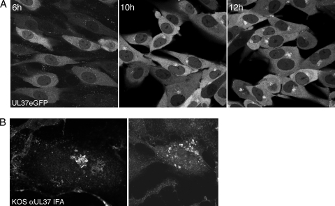
FIG. 3.
UL37eGFP localizes to a juxtanuclear site. (A) HFT cell monolayers (6 × 10⁵ cells per chamber slide) were infected with K37eGFP2 at an MOI of 10 PFU/cell and the cells visualized with a Zeiss LSM 510 Meta confocal microscope at 6, 10, and 12 h postinfection. The objective lens was 63×. (B) TIME cell monolayers were infected with KOS at an MOI of 10 PFU/cell. The cells were fixed at 15 h postinfection, stained with antibody to UL37 (780), and imaged by confocal analysis.

