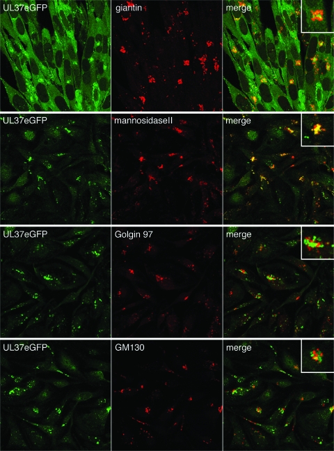
FIG. 4.
UL37eGFP localizes with giantin and mannosidase II but not with GM130 or Golgin 97. HFT or TIME cell monolayers were infected with K37eGFP2 at an MOI of 10 PFU/cell. The cells were fixed at 12 h (HFT) or 16 h (TIME) after infection and stained with antibodies to giantin (HFT cells), mannosidase II, Golgin 97, and GM130 (TIME cells). Images were collected using a confocal microscope. The GFP signal (pseudocolored green) was merged with the Cy3 signal (pseudocolored red) to determine the colocalization of the two colors and thus the two proteins. The objective lens was 63×.

