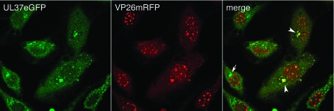
FIG. 5.
UL37eGFP traffics to the Golgi complex before capsids are transported to the same site. TIME cell monolayers in chamber slides were infected with K37eGFP/26R at an MOI of 10 PFU/cell and the cells visualized live with a confocal microscope at 16 h postinfection. White arrowheads indicate the colocalization of the two colors, and the arrow indicates the localization of UL37eGFP in the Golgi complex before transport of capsids to this site. The objective lens was 63×.

