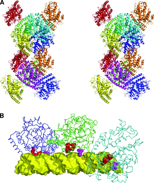
FIG. 4.
Assembly of σA molecules in the crystal and model of dsRNA binding. (A) Stereogram of the dodecameric assembly of σA in the crystallographic asymmetric unit. Monomers are shown in cartoon format and colored differently (two yellow, two orange, two red, two green, two blue, one magenta, and one cyan). (B) Model of a σA trimer interacting with a 36-bp canonical A-form dsRNA (yellow). Residues implicated in RNA binding, Arg155 and Arg273, are shown in red; Gln305 is shown in magenta.

